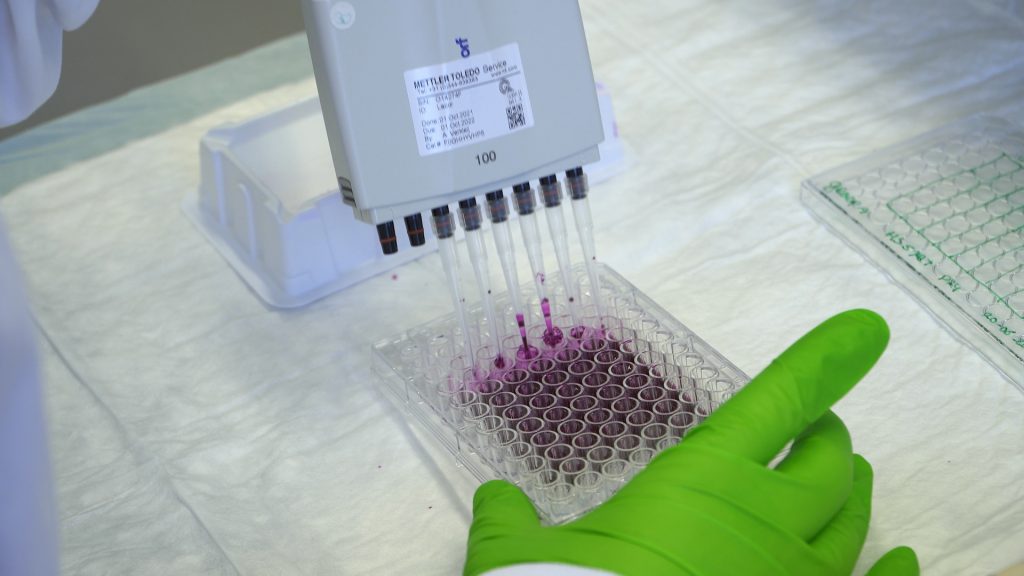
This video explains chemical and antibody staining of cells and how to execute the experimental steps.Visualization of proteins is accomplished by antibody staining.
Above you can see your plate layout. Calculate the volume needed of the first antibody stock and the volume of blocking solution. You have plated 6 wells per conditions, however you will only stain 2 wells per conditions (n = 2). The dilution factor is 1:2000 and each well can hold 100 µL of liquid. On top of the amount that you need to fill all wells, add 2 wells of volume to your calculation to compensate for pipetting errors.
We need µL of the first antibody (minval=0.9, maxval=0.9)
We need µL of the blocking solution (minval=1790, maxval=1810)
When storing your samples/compound in a lab, it is important to write down the information of your sample on the package. What information should be written down on your first antibody solution tube:
Choose one or more options:
(Team) name (correct)
Date (correct)
Dilution factor (correct)
Second antibody name
First antibody name (correct)
Diluted in.. (correct)
Tube size
All cells exhibit F-actin filaments. Why is it then useful to label F-actin? Pick one
F-actin forms a part of the cytoskeleton of the cell. The cell uses this to move, so that cell movement can be visualized.
F-actin forms a part of the cytoskeleton of the cell. As this determines the cell shape it makes visualization of the individual cells possible. (correct)
All cells exhibit F-actin filaments, but when the cell dies these filaments are broken down. The cell cannot be visualized then anymore; so, it is a viability marker.
All cells exhibit F-actin filaments, but when the cell divides these change structure which can then be observed during imaging; so, it is a proliferation marker.
Copyright information
This video is created by Leiden Academic Centre for Drug Research (LACDR), Faculty of Science at Leiden University under a open Creative Commons Attribution-NonCommercial-ShareAlike 4.0 International License. When using this video in its original version please refer to www.labprep.video. When adapting the video, mention the source ‘adapted based on the original version that is created by the labprep.video team’. It is not allowed to use the video for commercial purposes without consultation with the creators. You can contact us via info@labprep.video.