Fixation and blocking for immunofluorescence
In this video you will learn how to fixate and block cells for immunofluorescence assay or chemical staining.
- Tineke Bijl
-
(0)
- 0 enrolled students
Description
Non-interactive video download link (mp4). No video available, watch on YouTube instead.
The script that has been used to create the video shown above can be downloaded as excel script file. No file available.
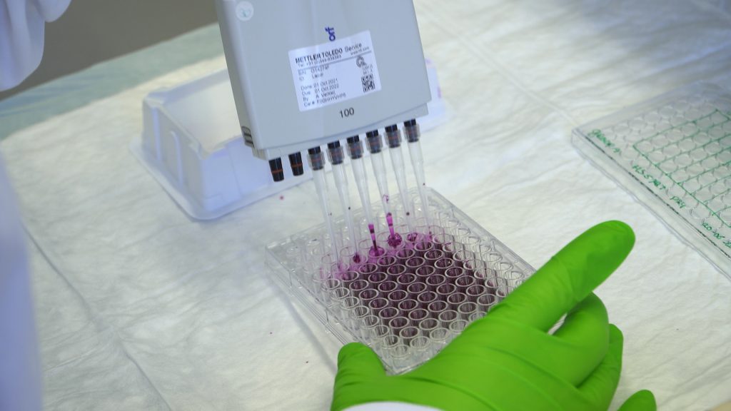
| 1 | Fixation Inspect your cells under the microscope. Write down: % confluency and any abnormalities that you observe. Select 2 wells per condition for antibody staining. |
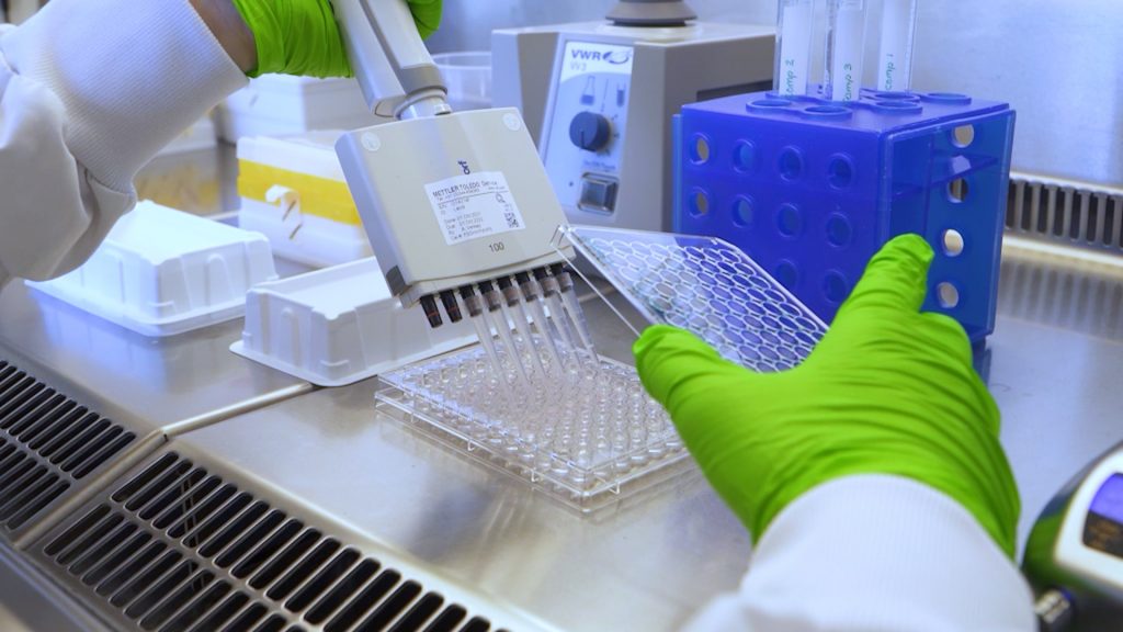
| 2 | Aspirate medium and wash the cells by adding 100 µl PBS per well, swirl the plate gently. |
| 3 | Aspirate PBS (flow cabinet) and add 100 µl PFA-Triton solution in each well. Incubate 15 minutes at room temperature. |
| 4 | Collect the Triton-X/PFA solution from the cells and discard in a 50 ml tube. Do not aspirate, since PFA is toxic. |
| 5 |
|
| 6 | Blocking Aspirate PBS from the cells. |
| 7 | Add 100 µl of blocking solution to each well with cells that you will stain. |
| 8 | Incubate 30 minutes at room temperature. |
PFA fixates the cells by crosslinking (covalent binding) specific amino acids. Why does this need to happen before the immunostaining?
Pick one
- Antibodies only bind to fixated proteins by binding to the crosslinks.
- Antibodies are broken down by living cells, so they must be fixated first.
- Cell fixation is needed to stop all protein synthesis and degradation at the same time point. (Correct)
- All the above statements are true.
PFA is not the only chemical that is added to the cells before immunostaining. Triton is also used, but what is the function of Triton?
Pick one
- Triton makes small holes in the cells so that the antibody can enter the cell.
- Triton is a catalysator for the fixation procedure.
- PFA is a solid matter and Triton is the liquid to dissolve it in
- Because of the Triton, the cells will not be stressed during fixation.
Why do you need to add blocking proteins before antibody staining?
Choose one or more options:
- To enhance background signal in the detection of the IF final product.
- To prevent the occurrence of false positives.
- To prevent the interaction between the antibody and the unspecific binding to the overall present proteins instead of binding to the target protein solely.
- To treat, i.e. ‘prepare’, the cells so that they can bind antibodies.
Which methods can be used to block samples prior to antibody staining?
Choose one or more options:
- Non-fat dry milk (correct)
- PBS-0.5% BSA solution (correct)
- Paraformaldehyde (PFA) in PBS
- I-Block with 0.1% of detergent (Tween 20 or Triton X-100) (correct)
- I-Block with 50% of detergent (Tween 20 or Triton X-100)

Download zip to import in LabBuddy. No file available.
Please make sure that your product exists and valid for this course
- Skill levelIntroduction video
- CategoryCell biology, Cytochemistry
Related videos
-
Free
Cell counting
Copyright information
This video is created by Leiden Academic Centre for Drug Research (LACDR), Faculty of Science at Leiden University under a open Creative Commons Attribution-NonCommercial-ShareAlike 4.0 International License. When using this video in its original version please refer to www.labprep.video. When adapting the video, mention the source ‘adapted based on the original version that is created by the labprep.video team’. It is not allowed to use the video for commercial purposes without consultation with the creators. You can contact us via info@labprep.video.